

3D “Spectracoustic” System: A Modular, Tomographic, Spectroscopic Mapping Imaging, Non-Invasive, Diagnostic System for Detection of Small Starting Developing Tumors like Melanoma
Fabrication of a combined acoustic microscopy transducer and infrared illumination probe permitting the simultaneous acquisition of the spectroscopic and the tomographic information. This work led to a new method named 3D spectracoustic tomographic mapping imaging. This probe provides the capability of high fidelity and precision registered information from the combined modalities, named spectracoustic information. This technology is also used for non invasive evaluation of cultural heritage objects.
In this project acoustic microscopy, infrared reflectance spectroscopy and imaging have been combined in order to study the structure of various types of small tumor lesions, like melanoma skin cancer. The tomographic images from a scanned region of interest (ROI) of the object are acquired using acoustic microscopy while the distribution of the materials in the ROI is acquired using infrared reflectance spectroscopy. Using the acquired spectra, the 3D segmentation – clustering of the spectra dataset can be derived in order to result in high resolution rendering of the internal structure of the object under investigation. The clusters consist of spectra that have similar characteristics — i.e., similar chemical composition. The spatial distribution of such clusters can be illustrated in pseudocolor images, in which each pixel is colored according to its cluster membership. Such mapping images convey information about the spatial distribution of the chemical substances in an object. A modular infrared spectroscope (Bruker ALPHA) in reflectance mode with a specially designed illumination area was used, after various trials, in order to acquire the array of spectra as well as a custom acoustic microscope for the acquisition of the tomographic data. The spectral area covered by spectroscope is 7500-375cm-1 (1.3-26μm). The wavelengths that are used ensure the penetration of the radiation in deeper layers. Furthermore, a control (CNC) system, which is driven by a software that is specially developed for the scanning of the object with the probes of all the modalities (acoustic microscopy, infrared reflectance spectroscopy), has been applied. Thus, the mapping images that are produced by clustering the acquired spectra in specific wavelength bands of infrared spectra can provide stratigraphic information, i.e., images that convey information of the distribution of substances or biochemical changes from deeper layers.
The various transducers were designed based on the ultrasonic of high frequency wave propagation simulation in melanoma simulating structure using Finite Difference Time Domain technique.
The system optical adaptation for the illumination area was designed based on simulation of electromagnetic waves propagation in melanoma simulated structure using Finite Integration technique.
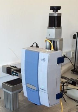
The infrared systems mounted on the XYZ moving stages (CNC) system 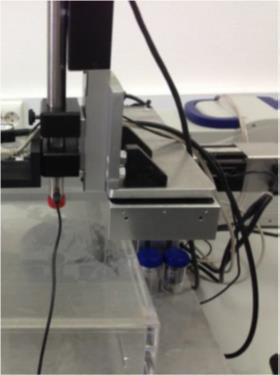
The acoustic microscopy transducer mounted on the XYZ moving stages (CNC) system
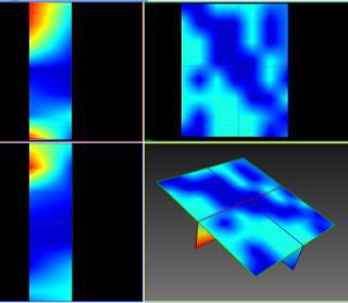
Spectroscopic mapping tomography 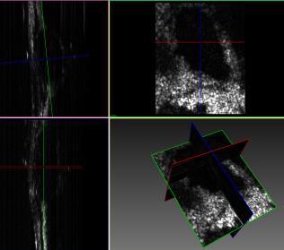
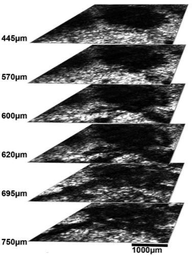
Acoustic micro-tomography
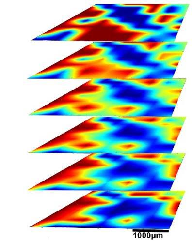
Unsupervised machine learning algorithms are applied for hierarchical clustering in the various wavelength areas chosen resulting to multispectral mapping images
The clustering images derived by infrared modality are registered on the tomographic images.
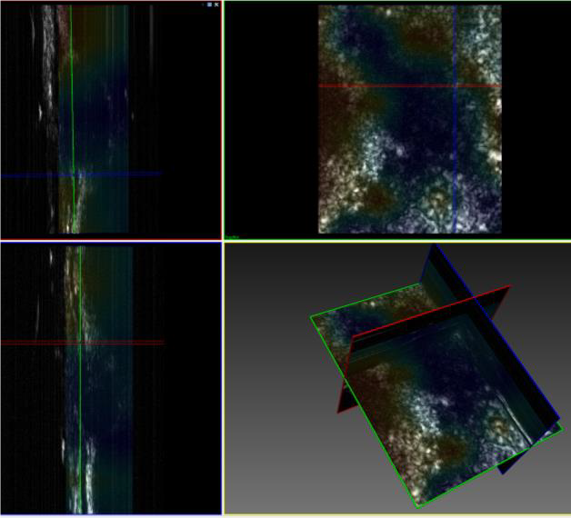
This registration provides the user a high fidelity information of the distribution of the materials in the 3D structure resulting in spectroscopic mapping imaging.
Results
This work led to a new method named “3D Spectracoustic Tomographic Mapping Imaging”. The current and the future work is related to the fabrication of a combined acoustic microscopy transducer and infrared illumination probe, permitting the simultaneous acquisition of the spectroscopic and the tomographic information.